45 eye diagram and labels
Human Eye Diagram, How The Eye Work -15 Amazing Facts of Eye The shark has even been used in human eye surgery! FACT 4 The length of our eyes are about 1 inch across and weigh about 0.25 ounce. FACT 5 Our eyeballs stay the same size forever but our nose and ears continue to grow. FACT 6 Eyes are the second most complex organ after the brain. Labelling the eye — Science Learning Hub In this activity, students use online or paper resources to identity and label the main parts of the human eye. By the end of this activity, students should be able to: identify the main parts of the human eye. describe the functions of the different parts of the human eye. Download the Word file (see link below).
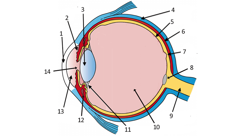
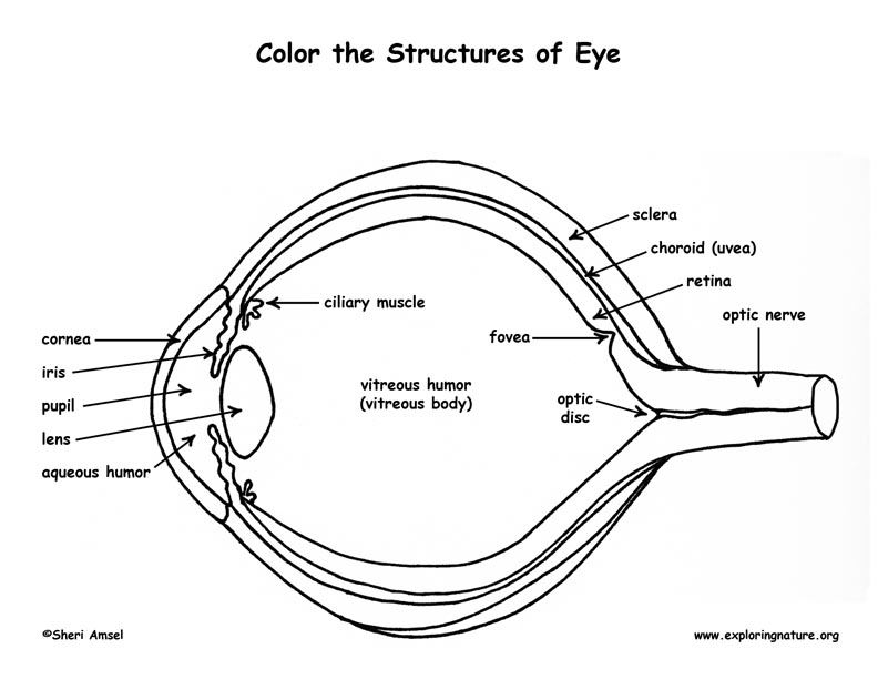
Structure and Functions of Human Eye with labelled Diagram The internal components of an eye are: Lens: It is a transparent, biconvex, lens of an eye. The lens is attached to the ciliary body by ligaments. The lens along with the cornea refracts light so that it focuses on the retina. Retina: It is the innermost layer of the eye. It is light sensitive and acts as a film of a camera.

Eye diagram and labels
Eye Diagram With Labels and detailed description - BYJUS A brief description of the eye along with a well-labelled diagram is given below for reference. Well-Labelled Diagram of Eye The anterior chamber of the eye is the space between the cornea and the iris and is filled with a lubricating fluid, aqueous humour. The vascular layer of the eye, known as the choroid contains the connective tissue. Labeled Eye Diagram | Science Trends What you want to interpret as a major part of the human eye is somewhat up to the individual, but in general there are seven parts of the human eye: the cornea, the pupil, the iris, the lens, the vitreous humor, the retina, and the sclera. Let's take a closer look at each of these components individually. The Cornea eye diagram with label eye labeled diagram anatomy human medical 11e3. Color Temperature hyperphysics.phy-astr.gsu.edu. chromaticity cie astr gsu phy hyperphysics engenharia representation blackbody 1931 determines incandescent. Human Eye: Anatomy, Parts And Structure - Online Biology Notes . eye anatomy human parts structure biology ...
Eye diagram and labels. Label Parts of the Human Eye Parts of the Eye Select the correct label for each part of the eye. The image is taken from above the left eye. Click on the Score button to see how you did. Incorrect answers will be marked in red. Labelling the eye — Science Learning Hub In this interactive, you can label parts of the human eye. Use your mouse or finger to hover over a box to highlight the part to be named. Drag and drop the text labels onto the boxes next to the eye diagram If you want to redo an answer, click on the box and the answer will go back to the top so you can move it to another box. Diagram of the Eye - Home - Lions Eye Institute To understand the eye and its functions, it's important to understand how the eye works, see below diagrams for both the external eye and the internal eye. The External Eye Instructions Click the parts of the eye to see a description for each. Hover the diagram to zoom. The Internal Eye Instructions Anatomy of the eye: Quizzes and diagrams | Kenhub One of our favorite ways to get to grips with all of the parts of the eye is by utilizing labeled diagrams. On a diagram of the eye, we can see all of the relevant structures together on one image. This helps us to understand how each one is situated and related to the other. Labeled diagram of the eye
Labelled Diagram of Human Eye, Explanation and Function - VEDANTU Labeled Diagram of Human Eye The eyes of all mammals consist of a non-image-forming photosensitive ganglion within the retina which receives light, adjusts the dimensions of the pupil, regulates the availability of melatonin hormones, and also entertains the body clock. 6,782 Human eye diagram Images, Stock Photos & Vectors - Shutterstock Find Human eye diagram stock images in HD and millions of other royalty-free stock photos, illustrations and vectors in the Shutterstock collection. Thousands of new, high-quality pictures added every day. Labeled Eye Diagram | Eye anatomy diagram, Eye anatomy, Diagram of the eye AS-IS: This vibrant 20" x 26" (55 x 61 cm) exam-room anatomy poster shows cross section of The Eye. It also provides lateral and superior view of the eye and shows the visual field. Anterior chamber angle, eyelashes, tear ducts, cornea, lens, retina, fundus and the macula lutea are illustrated. A good optometrist does more than issue ... Eye labeling Diagram | Quizlet Eye labeling STUDY Learn Flashcards Write Spell Test PLAY Match Gravity Created by csatNotaro Terms in this set (12) Retina the light-sensitive inner surface of the eye, containing the receptor rods and cones plus layers of neurons that begin the processing of visual information Eyeball organ of vision Sclera white of the eye Retinal blood vessels
Anatomy of the Eye Diagrams for Coloring/Labeling, with Reference and ... This printable contains 13 clear and simple cross sectional diagrams of the human eye. They photocopy well and are great for use as a labeling and coloring exercise for your students. The core eye anatomy diagram, designed as the labeling exercise, has a fully colored and labeled reference chart to go with it. Labeled Eye Diagram | Human eye diagram, Eye anatomy, Diagram of the eye Labeled Eye Diagram. Find this Pin and more on Legume by Nicole Young. Eye Anatomy Diagram. Human Eye Diagram. Diagram Of The Eye. Human Anatomy Drawing. Human Anatomy And Physiology. Anatomy Organs. Brain Anatomy. Eye diagram basics: Reading and applying eye diagrams - EDN Eye diagrams provide instant visual data that engineers can use to check the signal integrity of a design and uncover problems early in the design process. Used in conjunction with other measurements such as bit-error rate, an eye diagram can help a designer predict performance and identify possible sources of problems. Also see : eye diagram with labels eye diagram with labels Human Skeleton Back No Text No Color Clip Art at Clker.com - vector. 9 Pics about Human Skeleton Back No Text No Color Clip Art at Clker.com - vector : Muscles of the Human Eyeball | ClipArt ETC, Parts of an eye - ESL worksheet by step2eternity and also Human Body Anatomy Basics No Lines Clip Art at Clker.com - vector clip.
PDF Parts of the Eye - National Eye Institute | National Eye Institute Eye Diagram Handout Author: National Eye Health Education Program of the National Eye Institute, National Institutes of Health Subject: Handout illustrating parts of the eye Keywords: parts of the eye, eye diagram, vitreous gel, iris, cornea, pupil, lens, optic nerve, macula, retina Created Date: 12/16/2011 12:39:09 PM

anatomy, eye diagram to label | Kids science experiments/ crafts/school ideas | Pinterest | Eyes ...
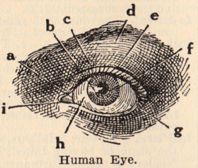
The Eyes (Human Anatomy): Diagram, Optic Nerve, Iris, Cornea ... - WebMD The front part (what you see in the mirror) includes: Iris: the colored part. Cornea: a clear dome over the iris. Pupil: the black circular opening in the iris that lets light in. Sclera: the ...
The Eye - diagram to label | Teaching Resources File previews. pdf, 2.94 MB. Diagram of eye with key words to use in labelling it. Tes classic free licence.
Eye Anatomy: 16 Parts of the Eye & Their Functions The following are parts of the human eyes and their functions: 1. Conjunctiva The conjunctiva is the membrane covering the sclera (white portion of your eye). The conjunctiva also covers the interior of your eyelids. Conjunctivitis, often known as pink eye, occurs when this thin membrane becomes inflamed or swollen.
diagram of eye with labels 32 Eye Diagram To Label - Labels Database 2020 ardozseven.blogspot.com. Pin On Examples Printable Label Templates . quizlet slidedocnow spinal cord. Chapter Two . brain sagittal human section anatomy labeled diagram cat robotspacebrain plane physiology drawing cut parts midsagittal study medical cranial diagrams ...
PDF Eye Anatomy Handout - National Eye Institute of light entering the eye. Lens: The lens is a clear part of the eye behind the iris that helps to focus light, or an image, on the retina. Macula: The macula is the small, sensitive area of the retina that gives central vision. It is located in the center of the retina. Optic nerve: The optic nerve is the largest sensory nerve of the eye.
The Eye Diagram: What is it and why is it used? The eye diagram is used primarily to look at digital signals for the purpose of recognizing the effects of distortion and finding its source. To demonstrate using a Tektronix MDO3104 oscilloscope, we connect the AFG output on the back panel to an analog input channel on the front panel and press AFG so a sine wave displays.
Parts Of The Eye Labeled Diagram Model And Their Function Parts of the eye-labeled diagram model are divided into three groups: the external outer layer, the middle layer, and the inner back layer. The outer layer is responsible for protecting the eye from environmental toxins and debris. The middle layer includes cells that allow light to enter and travel through the back layer to the retina.
Human Eye Diagram Without Labels - unlabeled eye diagram clipart best ... Human Eye Diagram Without Labels - 14 images - eye diagram images stock photos vectors shutterstock, the human eye activity bundle eye anatomy diagram eyeball diagram, plica semilunaris high resolution stock photography and images alamy, eye with labels clip art at vector clip art online royalty,
Eye diagram labeled - Healthiack Eye diagram labeled This summary post is displaying Eye diagram labeled … The eyes are responsible for our sense of sight or vision. Any disruption of the anatomy and physiology of the eyes and their supporting structures can cause vision impairment. Some of the ocular disorders or medical conditions affecting the eyes include the following:
eye diagram with label eye labeled diagram anatomy human medical 11e3. Color Temperature hyperphysics.phy-astr.gsu.edu. chromaticity cie astr gsu phy hyperphysics engenharia representation blackbody 1931 determines incandescent. Human Eye: Anatomy, Parts And Structure - Online Biology Notes . eye anatomy human parts structure biology ...
Labeled Eye Diagram | Science Trends What you want to interpret as a major part of the human eye is somewhat up to the individual, but in general there are seven parts of the human eye: the cornea, the pupil, the iris, the lens, the vitreous humor, the retina, and the sclera. Let's take a closer look at each of these components individually. The Cornea

Human Eye Diagram Labeled - Health, Medicine and Anatomy Reference Pictures | School | Pinterest ...
Eye Diagram With Labels and detailed description - BYJUS A brief description of the eye along with a well-labelled diagram is given below for reference. Well-Labelled Diagram of Eye The anterior chamber of the eye is the space between the cornea and the iris and is filled with a lubricating fluid, aqueous humour. The vascular layer of the eye, known as the choroid contains the connective tissue.



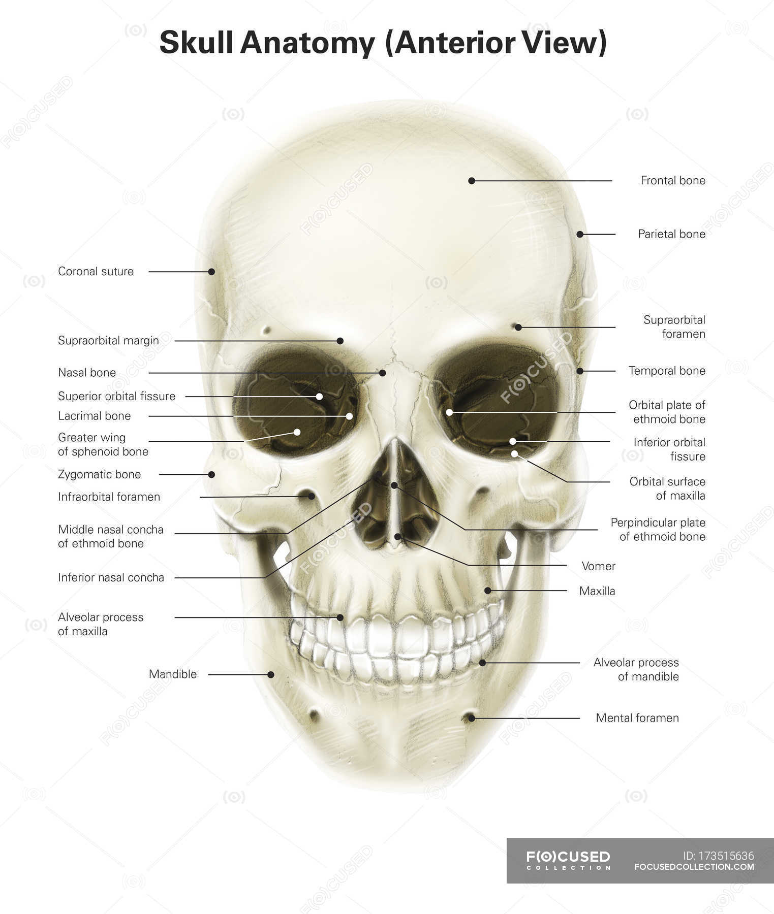
Post a Comment for "45 eye diagram and labels"