44 simple microscope diagram with labels
Compound Microscope Parts, Functions, and Labeled Diagram Compound Microscope Definitions for Labels. Eyepiece (ocular lens) with or without Pointer: The part that is looked through at the top of the compound microscope. Eyepieces typically have a magnification between 5x & 30x. Monocular or Binocular Head: Structural support that holds & connects the eyepieces to the objective lenses. Draw a neat labeled diagram for the formation of an image in a simple ... A simple microscope is a convex lens where in the image is kept between the focus of the lens so that the image formed is virtual and magnified. While drawing make sure the symmetry is taken into consideration. Complete step by step answer: Here is the labeled diagram outlining a simple microscope. To draw the ray diagram, place the object ...
A Study of the Microscope and its Functions With a Labeled Diagram ... May 21, 2019 - To better understand the structure and function of a microscope, we need to take a look at the labeled microscope diagrams of the compound and electron microscope. These diagrams clearly explain the functioning of the microscopes along with their respective parts.

Simple microscope diagram with labels
Labeling the Parts of the Microscope | Microscope World Resources Labeling the Parts of the Microscope. This activity has been designed for use in homes and schools. Each microscope layout (both blank and the version with answers) are available as PDF downloads. You can view a more in-depth review of each part of the microscope here. Simple Microscope - Parts, Functions, Diagram and Labelling Parts of the optical parts are as follows: Mirror - A simple microscope has a plano-convex mirror and its primary function is to focus the surrounding light on the object being examined. Lens - The biconvex lens is placed above the stage and its function is to magnify the size of the object being examined. Label the Microscope Diagram | Download Scientific Diagram In the study of antibiogram, all isolates were shown 100% sensitive to streptomycin, ciprofloxacin and chloramphenicol. Maximum 41% isolates were shown resistant to co-trimethaxozole whereas 30% ...
Simple microscope diagram with labels. Label Microscope Diagram - EnchantedLearning.com Using the terms listed below, label the microscope diagram. arm - this attaches the eyepiece and body tube to the base. base - this supports the microscope. body tube - the tube that supports the eyepiece. coarse focus adjustment - a knob that makes large adjustments to the focus. diaphragm - an adjustable opening under the stage, allowing ... Microscope Types (with labeled diagrams) and Functions A compound microscope: Is used to view samples that are not visible to the naked eye. Uses two types of lenses - Objective and ocular lenses. Has a higher level of magnification - Typically up to 2000x. Is used in hospitals and forensic labs by scientists, biologists and researchers to study micro organisms. Compound microscope labeled diagram. Microscope Poster - Diagram with Labels | Teach Starter A poster containing a diagram with labels showing the key parts of a microscope. In Science it is important that students know how to use a variety of tools when conducting scientific experiments and inquiry. This poster focuses on the microscope and highlights its key parts. Print on tabloid paper to display around your school's science lab ... Labeling Microscope Worksheet | Teaching Resources File previews. docx, 300.56 KB. A straightforward worksheet in which students are required to identify the parts of a basic microscope. Tes classic free licence.
Parts of a microscope with functions and labeled diagram Q. Differentiate between a condenser and an Abbe condenser. Ans. Condensers are lenses that are used to collect and focus light from the illuminator into the specimen. They are found under the stage next to the diaphragm of the microscope. They play a major role in ensuring clear sharp images are produced with a high magnification of 400X and above. Parts of a Simple Microscope - Labeled (with diagrams) image 5: A modern simple microscope with the different parts labeled. image source: laboratoryinfo.com The optical parts of a simple microscope are centered on the specimen - lighting, and magnification. Types of Microscopes: Definition, Working Principle, Diagram ... Where, D is the least distinct vision; F is the focal length of the convex lens; Simple Microscope Diagram. Principle of Simple Microscope. The working principle of a simple microscope is that when a sample is placed within the focus of the microscope, a virtual, erect and magnified image is obtained at the least distance of distinct vision from the eye that is held at the lens. Simple Microscope Definition, Magnification, Parts And Uses Following are the parts of the simple microscope with their functions: Eyepiece: It is the lens that is used to study the samples and is placed at the top. It has a magnification of 10X to 15X. Base: This provides support to the microscope. Tube: This is used to connect the eyepiece to the objective lenses.
Compound Microscope- Definition, Labeled Diagram, Principle, Parts, Uses The optical microscope often referred to as the light microscope, is a type of microscope that uses visible light and a system of lenses to magnify images of small subjects. There are two basic types of optical microscopes: Simple microscopes. Compound microscopes. The term "compound" in compound microscopes refers to the microscope having ... Microscope Parts and Functions Microscope Parts and Functions With Labeled Diagram and Functions How does a Compound Microscope Work?. Before exploring microscope parts and functions, you should probably understand that the compound light microscope is more complicated than just a microscope with more than one lens.. First, the purpose of a microscope is to magnify a small object or to magnify the fine details of a larger ... Simple Microscope - Diagram (Parts labelled), Principle, Formula and Uses Simple microscope is a magnification apparatus that uses a combination of double convex lens to form an enlarged, erect image of a specimen. The working principle of a simple microscope is that when a lens is held close to the eye, a virtual, magnified and erect image of a specimen is formed at the least possible distance from which a human eye ... Simple Columnar Epithelium Labeled Diagram In humans, a simple (Simple columnar epithelium labeled at right, third from top.).A simple columnar epithelium is a columnar epithelium that is uni-layered. In humans, a simple columnar epithelium lines most organs of the digestive tract including the stomach, . This type of epithelium is adapted for secretion and/or absorption, and can also ...
The Wonderful Microworld: Cell Nucleus - Onion
Microscope Labeling - The Biology Corner Microscope Labeling. Shannan Muskopf May 31, 2018. This simple worksheet pairs with a lesson on the light microscope, where beginning biology students learn the parts of the light microscope and the steps needed to focus a slide under high power. The labeling worksheet could be used as a quiz or as part of direct instruction where students ...

Label Microscope Diagram - ClipArt Best
PDF Parts of a Microscope Printables - Homeschool Creations Label the parts of the microscope. You can use the word bank below to fill in the blanks or cut and paste the words at the bottom. Microscope Created by Jolanthe @ HomeschoolCreations.net. Parts of a eyepiece arm stageclips nosepiece focusing knobs illuminator stage objective lenses
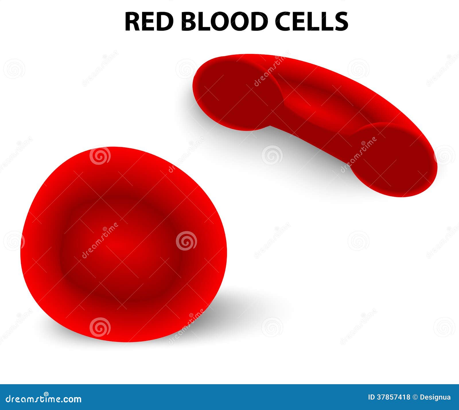
Red blood cells stock vector. Image of blood, healthy - 37857418
Compound Microscope Parts - Labeled Diagram and their Functions - Rs ... The term "compound" refers to the microscope having more than one lens. Basically, compound microscopes generate magnified images through an aligned pair of the objective lens and the ocular lens. In contrast, "simple microscopes" have only one convex lens and function more like glass magnifiers. [In this figure] Two "antique ...

Q14 Draw a large diagram of an animal cell as seen through an electron microscope. Label the ...
Label the microscope — Science Learning Hub Use this interactive to identify and label the main parts of a microscope. Drag and drop the text labels onto the microscope diagram. eye piece lens: The lens you look through - normally 10x or 15x magnification. eye piece lens. coarse focus adjustment: Moves the lens up or down and adjusts focus. coarse focus adjustment.
Print Anatomy Midterm 2 flashcards | Easy Notecards
Parts of a Microscope Labeling Activity - Storyboard That Knowing the names of the different parts of the microscope is essential to be able to use one properly. Create a poster that labels the parts of a microscope and includes descriptions of what each part does. Click "Start Assignment". Use a landscape poster layout (large or small). Search for a diagram of a microscope.
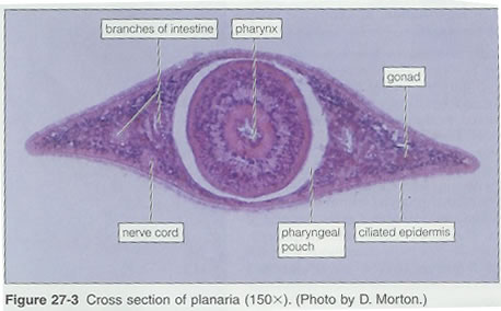
Flatworms
Microscope, Microscope Parts, Labeled Diagram, and Functions Revolving Nosepiece or Turret: Turret is the part of the microscope that holds two or multiple objective lenses and helps to rotate objective lenses and also helps to easily change power. Objective Lenses: Three are 3 or 4 objective lenses on a microscope. The objective lenses almost always consist of 4x, 10x, 40x and 100x powers. The most common eyepiece lens is 10x and when it coupled with ...

All Saints Online
Parts of the Microscope with Labeling (also Free Printouts) 5. Knobs (fine and coarse) By adjusting the knob, you can adjust the focus of the microscope. The majority of the microscope models today have the knobs mounted on the same part of the device. Image 5: The circled parts of the microscope are the fine and coarse adjustment knobs. Picture Source: bp.blogspot.com.

31 best images about Histology - GI - Layers, Junctions and Miscellaneous on Pinterest
Simple Microscope - Definition, Types, Working Principle & Formula Compound Microscope. 1. Simple microscope comprises a biconvex lens used as a magnifying glass. Compound microscope comprises 2 or more convex lenses where one lens is the eyepiece and the other one is the objective lens. 2. Natural light is the source to see the object. An illuminator is a source to see the object. 3.

www.timvandevall.com wp-content uploads Labeled-Microscope-Diagram.jpg | ชีววิทยาศาสตร์, ห้อง ...
Microscope labeled diagram - SlideShare Microscope labeled diagram 1. The Microscope Image courtesy of: Microscopehelp.com Basic rules to using the microscope 1. You should always carry a microscope with two hands, one on the arm and the other under the base. 2. You should always start on the lowest power objective lens and should always leave the microscope on the low power lens ...

microscope labeled microscope worksheet labeling sc 1 st template entrancing labelling - Top ...
Simple Microscope - Definition, Diagram, FAQs Define Microscope. Simple Microscope Definition: A Simple Microscope meaning is used to see a magnified image of an object. Antonie Van Leeuwenhoek, a Dutchman, invented the first simple microscope, consisting of a single powerful magnetic lens that rotates to detect tiny freshwater insects. It is composed mainly of light microscopes.

Microscope Diagram Unlabeled - Micropedia
Microscope Drawing And Label - Painting Valley All the best Microscope Drawing And Label 33+ collected on this page. Feel free to explore, study and enjoy paintings with PaintingValley.com ... Microscope Diagram L... 1200x927 1 0. Like JPG. Microscopic Drawing ... 791x1024 1 0. Like JPG. Draw A Large Diagram... 960x720 1 0. Like JPG. ... Simple Microscope Drawing. Microscope Drawing ...

Porifera diagram | Science biology, Arthropods, Plant science
Label the Microscope Diagram | Download Scientific Diagram In the study of antibiogram, all isolates were shown 100% sensitive to streptomycin, ciprofloxacin and chloramphenicol. Maximum 41% isolates were shown resistant to co-trimethaxozole whereas 30% ...

Amoeba Cell Diagram
Simple Microscope - Parts, Functions, Diagram and Labelling Parts of the optical parts are as follows: Mirror - A simple microscope has a plano-convex mirror and its primary function is to focus the surrounding light on the object being examined. Lens - The biconvex lens is placed above the stage and its function is to magnify the size of the object being examined.
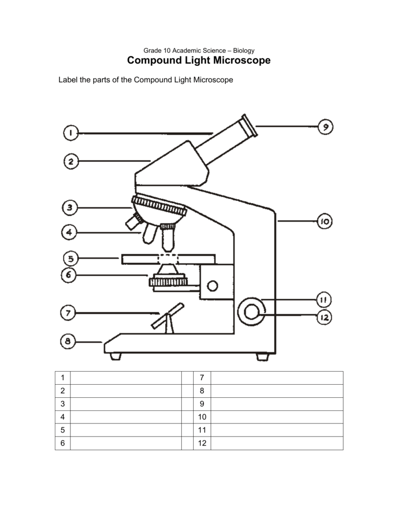
33 Microscope Diagram To Label - Labels Database 2020
Labeling the Parts of the Microscope | Microscope World Resources Labeling the Parts of the Microscope. This activity has been designed for use in homes and schools. Each microscope layout (both blank and the version with answers) are available as PDF downloads. You can view a more in-depth review of each part of the microscope here.
Post a Comment for "44 simple microscope diagram with labels"