41 muscle fiber model with labels
10.2 Skeletal Muscle - Anatomy & Physiology Figure 10.2.2 - Muscle Fiber: A skeletal muscle fiber is surrounded by a plasma membrane called the sarcolemma, which contains sarcoplasm, the cytoplasm of muscle cells. A muscle fiber is composed of many myofibrils, which contain sarcomeres with light and dark regions that give the cell its striated appearance. The Sarcomere Definition, Structure, Function and Quiz - Biology Dictionary Myosin is a thick fiber with a globular head, and actin is a thinner filament that interacts with myosin when we flex. Depicted is a basic illustration of skeletal muscle's underlying components, down to the sarcomere. Sarcomere structure. When viewed under a microscope, muscle fibers of varied lengths are organized in a stacked pattern.
PDF Anatomy & Physiology - Truckee Meadows Community College 15a. Smooth Muscle Cells 15b. Hyaline Top 15c. Part of the pushed out cell body Model KS 4 shows additionally… 16. Krause's endbulbs - with nerve coils in connective tissue cover 17. Ruffini's corpuscles -plexus of bare nerve fiber in connective tissue cover in the corium (dermis) and subcutis (hypodermis) 18.

Muscle fiber model with labels
OpenSim Models - OpenSim Documentation - Global Site Muscles typically include muscle activation and contraction dynamics and their own states (for example activation and muscle fiber length). The control values are typically bounded excitations (ranging from 0 to 1) which lead to a change in activation and then force. Below is an example of a muscle model, as described by Thelen (2003), from an ... Muscles Labeling - The Biology Corner Muscles Labeling. Shannan Muskopf November 11, 2020. This activity is aligned to my anatomy and physiology curriculum where students study the structure and function of muscle tissues. This has been a challenging topic to cover remotely because I can't use traditional models. Typically, I would use straws and rubber bands to model fascicles ... Sarcomere (Muscle) Coloring - Pinterest More on the elements of the flesh (sarco): Sarcolemma- plasma membrane of a muscle fiber, remember this has tube-like infoldings called transverse (T) tubules. Sarcoplasm- cytoplasm of muscle fiber that's occupied by myofibrils, glycogen, and myoglobin. Sarcomere- the contractile unit, a segment of myofibril from Z disc to the next Z disc.
Muscle fiber model with labels. quizlet.com › 304727113 › muscle-fiber-model-flash-cardsMuscle Fiber Model Flashcards | Quizlet Start studying Muscle Fiber Model. Learn vocabulary, terms, and more with flashcards, games, and other study tools. Search. ... Neuromuscular Junction labels (Ch.7) just numbers. 10 terms. 8A: NEURON MODEL. 26 terms. ... Neuron model. 7 terms. arm (upper arm) muscles. 6 terms. Muscles of the Lower Arm & Wrist. Features. Quizlet Live. Labeled Sarcomere Diagram The thin filaments Look at the diagram above and realize what happens as a muscle contracts. As will soon be described, the functional unit of a skeletal muscle fiber is the sarcomere, a highly organized arrangement of the contractile myofilaments actin .Play this quiz called Label the Sarcomere and show off your skills. Deep Learning Models to Characterize Smooth Muscle Fibers in ... Thick muscle layer underneath lamina propria is the muscularis propria (MP), also known as detrusor muscle. The final layer is the serosa/adventitia that covers the bladder dome. The MM and MP are the two types of smooth muscle fibers seen in the urinary bladder. The MM is composed of several thin layers of muscle fibers, often showing How to Build a Muscle Model | eHow When studying the muscular system, it can be helpful to look upon a functioning model of a muscle, which can help you visualize how a muscle works. The human body is comprised of bones, tissues and muscles that enable the body to move in a wide range of motions. When studying the muscular system, it can be helpful to look upon a functioning ...
Muscle fiber model Quiz - PurposeGames.com This is an online quiz called Muscle fiber model There is a printable worksheet available for download here so you can take the quiz with pen and paper. Your Skills & Rank Total Points 0 Get started! Today's Rank -- 0 Today 's Points … us.vwr.com › store › product3B Scientific® MICROanatomy™ Muscle Fiber Model - VWR This model features muscle tissue magnified by 10,000x.Depicting functions and locations, the human biology models allow individuals a more comprehensive understanding of internal organs and systems. Blood movement, oxygen transport, muscle constriction, nerve firing, joint movement, and bone formation are brought to life in these incredibly detailed and accurate representations. Musculature ... › en › libraryLearn all muscles with quizzes and labeled diagrams | Kenhub Apr 26, 2022 · Human body muscle diagrams. Muscle diagrams are a great way to get an overview of all of the muscles within a body region. Studying these is an ideal first step before moving onto the more advanced practices of muscle labeling and quizzes. If you're looking for a speedy way to learn muscle anatomy, look no further than our anatomy crash courses . Student Exploration Muscles and Bones Gizmo Vocabulary: actin, biceps, cartilage, contract, extend, fast twitch fiber, flex, fulcrum, humerus, lever, ligament, muscle fiber ... Gizmo, you will learn about the anatomy of the arm. You will build an arm model, then test its ability to lift a dumbbell and throw a ball. ... Select Hide muscles. Click Play. Describe what the arm bones are ...
PDF THE MUSCULAR SYSTEM - University of Cincinnati receptors on the muscle fiber membrane. The proteins inside muscle fibers are organized into long chains that can interact with each other, reorganizing to shorten and relax. When acetylcholine reaches receptors on the membranes of muscle fibers, membrane channels open and the process that contracts a mastering bio questions- exam 3- bio 132 Flashcards - Quizlet Most of the neurons in the human brain are. interneurons. The nucleus and most of the organelles in a neuron are located in the. cell body. The point of connection between two communicating neurons is called the. synapse. A neuron's nucleus is located in its _____. cell body. PDF ANATOMY OF THE MUSCULAR SYSTEM - Midland Independent School District the skeletal part that is moved when the muscle contracts (insertion). 1. Identify the connective tissue membrane that: (a) covers individual muscle fibers, (b) surrounds groups of skeletal muscle fibers (fascicles), and (c) covers the muscle as a whole. 2. Name the tough connective tissue cord that serves to attach a muscle to a bone. 3. Solved art A rag the labels onto the diagram to identify the - Chegg Science. Anatomy and Physiology. Anatomy and Physiology questions and answers. art A rag the labels onto the diagram to identify the parts of a cardiac muscle fiber Reset Help tubule of DELL.

Patellar reflex - wikidoc
Muscle Fiber Labeling Quiz - PurposeGames.com This is an online quiz called Muscle Fiber Labeling Quiz. There is a printable worksheet available for download here so you can take the quiz with pen and paper. Your Skills & Rank. Total Points. 0. ... Knee Joint Model Labeling Quiz 15p Image Quiz. Synovial Joint of Left Knee Labeling Quiz 10p Image Quiz. Blood Cells 7p Image Quiz ...
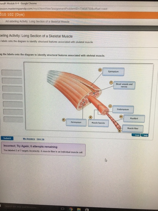
Drag The Labels Onto The Diagram To Identify Structural Features Associated With Skeletal Muscle ...
› health › muscle-fibersMuscle Fibers: Anatomy, Function, and More - Healthline May 12, 2020 · Takeaway. The muscular system works to control the movement of our body and internal organs. Muscle tissue contains something called muscle fibers. Muscle fibers consist of a single muscle cell ...
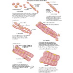
Development of Skeletal Muscle Fibers
Neurolemmocyte On Skeletal Muscle Model - GUWS Medical Use figure 18.1 of skeletal muscle tissue to locate the following features: skeletal muscle fiber (cell) nuclei striations (alternating light and dark) 3. Review a textbook section on structure of a skeletal muscle. 4. Study figures 18.2 and 18.3. 5. Examine the torso and locate examples of fascia, tendons, and aponeuroses.

75 best Anatomy & Physiology Stuff images on Pinterest | School, Anatomy and Human body
Muscle Fiber Model (Altay) Flashcards - Quizlet Muscle fiber model identifications Terms in this set (21) sarcolemma Identify the membrane. endomysium Identify the tissue layer. myofibril Identify the structure. thick myofilament …

The Humanengine book - HUMANENGINE
Muscle Fiber Model #1 - Ohio University - Anatomy Mar 26, 2019 · Muscle Fiber Model #1 - Ohio University - Anatomy & Physiology

The Contractile And Energetic Properties Of Skeletal Muscle Fibers - Smooth Muscle
DOCX Label the diagram of the skeletal muscle with the following terms ... Label the diagram of the muscle fiber below with the following terms: actin (thin filaments), neuromuscular junction, myofibril, myosin (thick filaments), sarcolemma, sarcomere, sarcoplasmic reticulum, T-tubule, Z-line. 3. Color the muscles of the head and neck in the diagrams below. 4. Color the muscles of the upper back and chest in the ...

Dbios Anatomical Model Micro Anatomy Of Muscle Fiber, For Medical, Size: 23x26x18cms, | ID ...
Muscle Charts of the Human Body - PT Direct PT Program Template. FREE Download. Make writing personal training programs easy with these custom designed exercise templates, and keep your clients focused and progressing.

Replika, Taman, Meubel, Jualan, Franchise, Pembantu, Pegawai, Mahasiswi, Guru, Gordyn, Jok ...
Solved 8 of 8 Review Learning Goal: To learn the parts - Chegg Anatomy and Physiology. Anatomy and Physiology questions and answers. 8 of 8 Review Learning Goal: To learn the parts of a smooth muscle fiber. Label the parts of a smooth muscle fiber. Part A Drag the labels onto the diagram to identify the parts of a smooth muscle fiber. Reset Help Intermediate filaments (desmin) Intermediate filaments.

V Ling: 08.10
Microscopic Structure Of Skeleton Muscles - Anatomy Notes MICROSCOPIC STRUCTURE OF SKELETON MUSCLES. The skeletal muscle fiber is known as enormous multinucleate cells and it develops through the fusion of each myoblast. These myoblasts are long, cylindrical structures with consistent size within the muscles. The size of myoblast may vary in different muscles from 10 to 100 µm in diameter, and from a ...

Muscle and Models on Pinterest
10.3 Muscle Fiber Contraction and Relaxation - OpenStax Relaxing skeletal muscle fibers, and ultimately, the skeletal muscle, begins with the motor neuron, which stops releasing its chemical signal, ACh, into the synapse at the NMJ. The muscle fiber will repolarize, which closes the gates in the SR where Ca ++ was being released. ATP-driven pumps will move Ca ++ out of the sarcoplasm back into the SR.

7 best Weight Training Class images on Pinterest | Muscles, Strength training and Weight training
Anatomy Models - Anatomical Models and Keys - NEOMED Muscle Fiber. Bone Structure. Liver Microanatomy . Heart and Diaphragm . Larynx . Base of Head . Small Liver Model . Liver Denoyer Model . Kidney Denoyer Model Disc/MRI Head . Smooth Muscle . Heart and Lung Larynx is missing . Brain Stem Enlarged 3X. Nerves of the Head. Brain Stem (SOMSO) - Detailed. Cranial Layers . Diencephalon. Ventricles ...

11 x 17 Post-It Anatomical Chart: HUMAN MUSCLE TYPES: Science Prints: Amazon.com: Industrial ...
Fluorophore‐labeled myosin‐specific antibodies simplify muscle‐fiber ... Skeletal muscles are frequently analyzed for composition of phenotypically distinct myofibers, as a functional determinant. We describe an improved myofiber phenotyping procedure, involving cryosecti...

Anatomy/Muscular System - Wiki - Scioly.org
SAC A&P Model Key - Muscular System - austincc.edu Muscular System. M1 - Muscled Arm. M2 - Muscle Leg. M3 - Female Muscle Figure. M4 - Microanatomy Muscle Fiber. M5 - Muscle Figure.

Muscle Fiber Types | Renaissance Man Journal
MUSCLE MODEL ACTIVITY GUIDE - Field Museum muscle model / The Machine inside: BioMechanics activity guide For stud E nt ACTIVITY – Exploring Muscle Size Most animals, including humans, are born with the exact number of …

Muscle Fiber Model Labeled | Muscle anatomy, Skeletal muscle anatomy, Physiology
PDF Muscle Physiology - ISD 2135 Maple River Schools / Homepage 2. Detail the functions of the muscle system. 3. Correctly label the parts of a myocyte (muscle cell) 4. Identify the levels of organization in a skeletal muscle from organ to myosin. 5. Explain how a muscle contracts utilizing the correct terminology of the sliding filament theory. 6. Contrast and compare cardiac and smooth muscle with ...

What Did You Do Today at School?: Muscle Contraction Modeling
Skeletal Muscle Fiber Model - Myofibrils - YouTube Jan 07, 2009 · This video was produced to help students of human anatomy at Modesto Junior College study our anatomical models.
Post a Comment for "41 muscle fiber model with labels"